Surface Plant Opals of Setaria viridis Seeds Correlate with Seed Dormancy
-
Inako Rina
Regional Field Science Research Center, Faculty of Agriculture, Shizuoka University, Shizuoka 422-8529, 835-1 Suruga-ku, Shizuoka, Japan
Inagaki Hidehiro

Regional Field Science Research Center, Faculty of Agriculture, Shizuoka University, Shizuoka 422-8529, 835-1 Suruga-ku, Shizuoka, Japan
| Received 25 Apr, 2025 |
Accepted 23 Jun, 2025 |
Published 25 Jun, 2025 |
Background and Objective: Plant opals, formed by the deposition of soluble silicic acid in plant cells, serve structural and defensive functions in leaves and stems. However, their role on seed surfaces remains unclear. This study investigates the structure and potential function of plant opals on the seeds of Setaria viridis, a species exhibiting seed dormancy. The objective was to determine whether these surface opals are associated with dormancy regulation. Materials and Methods: Seed surface morphology was analyzed using scanning electron microscopy. Comparative observations were made between S. viridis and S. italica (a non-dormant cultivated form). A standard cold and moist stratification treatment was applied to assess dormancy release. Observations focused on changes in seed coat structure, particularly the detachment or cracking of plant opals. No additional statistical tests were required for this morphological comparison. Results: A distinct parallel arrangement of plant opals was observed on the outer seed coat of S. viridis. These opals detached following dormancy-breaking treatment, initiating cracks in the seed coat. In contrast, S. italica exhibited pre-damaged or missing opal structures and lacked dormancy. These findings suggest a correlation between intact opal structures and seed dormancy, implying that opal detachment may facilitate water permeability and germination. Conclusion: The plant opals on the seed surface of S. viridis may contribute to seed dormancy by forming a physical barrier. Their structural disruption during dormancy-breaking treatments supports a role in regulating seed coat permeability. Further research is necessary to clarify the biochemical and mechanical processes involved.
| Copyright © 2025 Rina and Hidehiro. This is an open-access article distributed under the Creative Commons Attribution License, which permits unrestricted use, distribution, and reproduction in any medium, provided the original work is properly cited. |
INTRODUCTION
Setaria viridis is an annual Poaceae plant that performs C4 photosynthesis, similar to other major global crops such as maize, sorghum, and sugarcane. These major C4 crops are large and not suitable for cultivation in laboratories. However, S. viridis has the same NADP-ME type of photosynthesis subtype and excellent characteristics such as a small plant and genome size, making it suitable for use as a model C4 plant1,2. However, the seeds of S. viridis show deep dormancy and do not germinate uniformly, making it difficult to use S. viridis as a model plant3,4. Although chemical treatments, such as gibberellin and liquid smoke, are effective4,5, it remains challenging to achieve stable germination5. Thus, developing seed dormancy-breaking methods is important to utilize S. viridis as a model plant6-11.
One of the factors contributing to seed dormancy in S. viridis is the water-repellent structure of the outer coat12. In this study, the surface structure of the seed coat of S. viridis was carefully observed using an electron microscope, and the presence of plant opals on the seed surface was confirmed. Plant opals are characterized by the accumulation of soluble silicic acid in plant cells. In leaves and stems, plant opals function as skeletal tissue and protect the plant from grazing and disease13,14. By contrast, the role of plant opals on seeds is unknown. This study shows that plant opals on the seed surface may play a role in dormancy.
MATERIALS AND METHODS
Plant material: Setaria viridis seeds were collected from the Regional Field Science Research Center, Faculty of Agriculture, Shizuoka University, (6334.9052333N, 138.2721461E, Kariyado, Fujieda, Japan) in October, 2023. No germination was observed immediately after collection, confirming dormancy. Germinated seeds broken from dormancy by cold and wet treatment at 5°C for 4 weeks were prepared.
For comparison, prepared Setaria italica, a cultivated species derived from S. viridis, and confirmed that S. italica seeds were not dormant and had a high germination rate.
Electron microscope: A scanning electron microscope (Miniscope®TM3030Plus, Hitachi High-Tech Corporation Tokyo) was used to observe the fine structure. Twenty dormant seeds, 20 germinated seeds of S. viridis, and 20 ungerminated seeds of S. italica were observed using electron microscopy.
Statistical analysis: The germination rates were compared using the Bell Curve for Excel 5.0 (Social Survey Research Information Co., Ltd.) with Fisher’s exact test.
RESULTS AND DISCUSSION
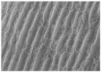
Immediately after the seed harvest, plant opals were observed as dumbbell-shaped and arranged in parallel on the outer coats of all dormant S. viridis seeds (Fig. 1).
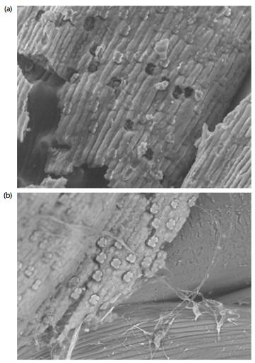
However, after the stratification treatment, the plant opals on the outer coat of the seeds peeled off. Cracks were observed to form on the outer coat of the seeds, starting from the peeled-off points in all germinated seeds (Fig. 2a-b); this suggests that the plant opals on the outer coat of the seeds affect the impermeability of the seed coat and the disappearance or damage of plant opals may trigger the breaking of dormancy.
Furthermore, in the case of S. italica, even in untreated seeds that had not undergone stratification, holes, cracks, and the loss of plant opals were observed in all ungerminated seeds. Unlike S. viridis, cultivated S. italica does not exhibit deep dormancy; it is speculated that this is because the outer coat shrinks during cultivation, causing the structure of the plant opals to be lost.
Recently, S. viridis has garnered attention as a model C4 plant1. However, deep seed dormancy of S. viridis limits its use as a model plant. Hence, to use S. viridis as a model plant, it is essential to develop a stable method to break dormancy6-11.
In general, storing Setaria seeds in the dark under refrigerated conditions is effective for breaking their dormancy5,13. However, this treatment requires several months to induce germination. Therefore, chemical treatment methods, such as liquid smoke treatment, gibberellic acid treatment14,15, and KNO3 treatment4, have been proposed to induce germination in a shorter period. However, it has been reported that the smoke liquid treatment harms seeds, resulting in a low germination rate5. In addition, the effect of any chemical treatments on seed germination requires several weeks and is unstable, and with these treatments4, it is relatively difficult to achieve consistent and uniform germination5.
|
|
Meanwhile, it has been suggested that the water-repellent structure of the outer skin may be related to dormancy in S. viridis16,17. The study demonstrated the possibility that plant opal acts as a water tap, destroying the water-repellent seed coat structure and triggering the breaking of dormancy. Current observations support the hypothesis that dormancy in S. viridis depends on the water-repellent structure of the seed coat.
Conversely, plant opals are characterized by the accumulation of soluble silicic acid in plant cells. Plant opals are physically stable and remain in the soil as microfossils even after the plant body dies. Therefore, plant opal analyses are mainly conducted in the field of archaeology, such as in vegetation surveys of ruins16,17. However, the role of opals in living plants remains unknown. It is thought that plant opals present in leaves and stems function as plant skeletal tissue and protection from feeding and disease. By contrast, the role of plant opals observed on seeds is unknown. In response to this issue, this report is the first to show that the phytoplankton on the seed surface may act as a water tap and be involved in breaking dormancy. In the future, a deeper understanding of the seed surface structure of S. viridis, particularly the plant opals on the seed coat, could provide an approach to breaking dormancy.
CONCLUSION
The plant opals on the outer coat of S. viridis seeds may act as water faucets, affecting seed dormancy. Their structural disruption during dormancy-breaking treatments plays a role in regulating seed coat permeability. Further research is needed to examine the role of plant opals by observing and analyzing the microstructure of the seed surface of S. viridis in three dimensions.
SIGNIFICANCE STATEMENT
There are many unknowns regarding the role of phytoliths in seeds. The results of this study revealed that the phytoliths on the seed surface of S. viridis act as a physical water tap, and the destruction of this tap may contribute to seed dormancy. The deep seed dormancy in S. viridis is considered a constraint when using it as a model plant, and the findings of this study may solve this problem. Furthermore, the observations on the role of the phytoliths in S. viridis in this study provide important insights into the role of phytoliths in seeds.
REFERENCES
- Li, P. and T.P. Brutnell, 2011. Setaria viridis and Setaria italica, model genetic systems for the Panicoid grasses. J. Exp. Bot., 62: 3031-3037.
- Brutnell, T.P., J.L. Bennetzen and J.P. Vogel, 2015. Brachypodium distachyon and Setaria viridis: Model genetic systems for the grasses. Annu. Rev. Plant Biol., 66: 465-485.
- Manthey, D.R. and J.D. Nalewaja, 1987. Germination of two foxtail (Setaria) species. Weed Technol., 1: 302-304.
- Sebastian, J., M.K. Wong, E. Tang and J.R. Dinneny, 2014. Methods to promote germination of dormant Setaria viridis seeds. PLoS ONE, 9.
- Acharya, B.R., S.R. Choudhury, A.B. Estelle, A. Vijayakumar, C. Zhu, L. Hovis and S. Pandey, 2017. Optimization of phenotyping assays for the model monocot Setaria viridis. Front. Plant Sci., 8.
- Muthamilarasan, M., V.S. Bonthala, R. Khandelwal, J. Jaishankar, S. Shweta, K. Nawaz and M. Prasad, 2015. Global analysis of WRKY transcription factor superfamily in Setaria identifies potential candidates involved in abiotic stress signaling. Front. Plant Sci., 6.
- Feng, Z.J., Z.S. Xu, J. Sun, L.C. Li and M. Chen et al., 2016. Investigation of the ASR family in foxtail millet and the role of ASR1 in drought/oxidative stress tolerance. Plant Cell Rep., 35: 115-128.
- Huang, P., H. Jiang, C. Zhu, K. Barry and J. Jenkins et al., 2017. Sparse panicle1 is required for inflorescence development in Setaria viridis and maize. Nat. Plants, 3.
- Feldman, M.J., R.E. Paul, D. Banan, J.F. Barrett and J. Sebastian et al., 2017. Time dependent genetic analysis links field and controlled environment phenotypes in the model C4 grass Setaria. PLoS Genet., 13.
- Liu, K., S. Qi, D. Li, C. Jin and C. Gao et al., 2017. TRANSPARENT TESTA GLABRA 1 ubiquitously regulates plant growth and development from Arabidopsis to foxtail millet (Setaria italica). Plant Sci., 254: 60-69.
- van Eck, J. and K. Swartwood, 2015. Setaria viridis. In: Agrobacterium Protocols, Wang, K., Springer, New York, ISBN: 978-1-4939-1695-5, pp: 57-67.
- Hidehiro, I. and S. Takumi, 2020. Effect of physical treatments on the promotion of seed germination in green and giant foxtails. Am. J. Agric. Biol. Sci., 15: 126-131.
- Fawe, A., M. Abou-Zaid, J.G. Menzies and R.R. Belanger, 1998. Silicon-mediated accumulation of flavonoid phytoalexins in cucumber. Biochem. Cell Biol., 88: 396-401.
- Rodrigues, F.Á., D.J. McNally, L.E. Datnoff, J.B. Jones and C. Labbé et al., 2004. Silicon enhances the accumulation of diterpenoid phytoalexins in rice: A potential mechanism for blast resistance. Phytopathology, 94: 177-183.
- Amini, V., F. Zaefarian and M. Rezvani, 2015. Interspecific variations in seed germination and seedling emergence of three Setaria species. Braz. J. Bot., 38: 539-545.
- Rovner, I., 1988. Macro- and micro-ecological reconstruction using plant opal phytolith data from archaeological sediments. Geoarchaeology, 3: 155-163.
- Rovner, I., 1971. Potential of opal phytoliths for use in paleoecological reconstruction. Quat. Res., 1: 343-359.
How to Cite this paper?
APA-7 Style
Rina,
I., Hidehiro,
I. (2025). Surface Plant Opals of Setaria viridis Seeds Correlate with Seed Dormancy. Research Journal of Seed Science, 18(1), 17-21. https://doi.org/10.3923/rjss.2025.17.21
ACS Style
Rina,
I.; Hidehiro,
I. Surface Plant Opals of Setaria viridis Seeds Correlate with Seed Dormancy. Res. J. Seed Sci 2025, 18, 17-21. https://doi.org/10.3923/rjss.2025.17.21
AMA Style
Rina
I, Hidehiro
I. Surface Plant Opals of Setaria viridis Seeds Correlate with Seed Dormancy. Research Journal of Seed Science. 2025; 18(1): 17-21. https://doi.org/10.3923/rjss.2025.17.21
Chicago/Turabian Style
Rina, Inako, and Inagaki Hidehiro.
2025. "Surface Plant Opals of Setaria viridis Seeds Correlate with Seed Dormancy" Research Journal of Seed Science 18, no. 1: 17-21. https://doi.org/10.3923/rjss.2025.17.21

This work is licensed under a Creative Commons Attribution 4.0 International License.